
A patient once asked me:
“If the PET scan is negative, doesn’t that mean it cannot be cancer?”
Unfortunately, not always.
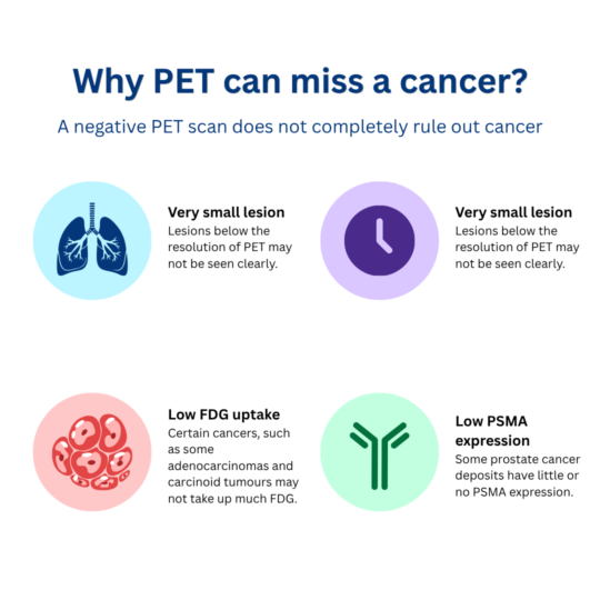
A PET-negative lung nodule can still sometimes be cancer. A negative PET scan can be reassuring, but it does not completely rule out cancer. Some abnormalities are too small to show clearly. Some cancers grow slowly or do not take up much of the radioactive tracer used for the scan.
That is why I never interpret a PET scan in isolation.
The CT appearance, whether an abnormality is changing over time, the patient’s previous cancers, blood tests and the clinical history can all be just as important.
One patient I recently reviewed demonstrated this particularly well.
Who is this article for?
This article may be helpful if:
- you have a PET-negative lung nodule that is being monitored;
- a lung nodule has grown despite a negative PET scan;
- you have prostate cancer and a rising PSA despite a negative PSMA PET;
- an unexpected abnormality was found elsewhere on your PET scan; or
- you are wondering why your doctors are still recommending further investigation after reassuring imaging.
The short answer
Yes, a PET-negative lung nodule can still be cancer.
FDG PET and PSMA PET are powerful tests, but neither detects every cancer. Small lesions and cancers with low tracer uptake can be missed.
If a nodule is enlarging on serial CT scans, that change may remain important even when PET imaging is negative. Current PET guidance also emphasises interpreting PET findings together with CT, previous imaging and the clinical context.
A complicated cancer history
This gentleman had previously been treated for prostate cancer.
He underwent prostate surgery in 2014, followed by radiotherapy, and later required further radiation treatment.
His PSA had subsequently risen to 19.4 ng/mL.
After radical prostatectomy, PSA would usually be expected to fall to an undetectable or very low level. A rising PSA therefore raises concern for recurrent prostate cancer, although the PSA alone cannot tell us where that recurrence is located.
He also had a previous history of multiple myeloma and had undergone a stem-cell transplant.
More recently, CT imaging showed several small nodules in his left lung. When the scans were compared over time, the nodules appeared to have increased slightly in size.
We therefore had several possibilities:
Was this recurrent prostate cancer? A new lung cancer? Something related to his previous myeloma? Or a completely unrelated process?
Why were two different PET scans performed?
The two scans were looking for different biological signals.
FDG PET
FDG PET uses a tracer similar to glucose. Many cancers use glucose more actively than surrounding tissues and therefore appear brighter or “hotter” on the scan.
PSMA PET
PSMA PET uses a tracer that binds to prostate-specific membrane antigen (PSA) , a protein expressed by many prostate cancer cells.
It is particularly useful when we are looking for prostate cancer that may have returned after previous treatment.
In this patient, however, neither tracer showed significant activity in the lung nodules.
That sounds reassuring.
But there was another piece of evidence we could not ignore:
the nodules appeared to be growing on CT.
| Test | What it helps show | What it may not tell us |
| CT scan | Size, shape, density and whether a nodule is growing | Whether the tissue is biologically active |
| FDG PET | Areas with increased glucose metabolism | Very small, slow-growing or low-FDG-uptake cancers can sometimes be missed |
| PSMA PET | Areas expressing PSMA, especially in prostate cancer | Some prostate cancer deposits with low PSMA expression |
| Biopsy | What the abnormal cells actually are | It may not always be safe, technically possible or necessary |
These tests answer different questions, which is why one reassuring result does not always settle the diagnosis.
Can a PET-negative lung nodule still be cancer?
Yes.
One systematic review and meta-analysis involving 1,301 pulmonary nodules found that FDG PET/CT had an overall sensitivity of about 82% for identifying malignant nodules.
Put simply, across those studies, FDG PET/CT detected about 82 in every 100 malignant lung nodules. In other words, some cancers were still PET-negative. The exact performance varies substantially according to the type and size of the nodule.
FDG PET can be less sensitive for:
- very small nodules;
- slow-growing cancers;
- some lung cancers that appear hazy or partly solid on CT; and
- certain tumour types that may take up relatively little FDG, including some adenocarcinomas and carcinoid tumours.
Small lesion size itself can reduce the apparent uptake because PET has limited spatial resolution.
This is why I would avoid saying:
“The PET scan is negative, therefore the nodule is benign.”
A better interpretation is:
“The nodule is not showing significant FDG activity, but we still need to interpret that result alongside its CT appearance and behaviour over time.”
Why growth on CT matters
A PET scan gives us information about biological activity at a particular point in time.
Serial CT scans tell us something different:
Is the nodule stable, disappearing or growing?
Growth does not automatically mean cancer. Infection, inflammation and other conditions can also change over time.
But documented growth can increase concern and may change whether surveillance, further imaging or tissue sampling is appropriate. Doctors consider what the nodule looks like on CT, how dense it is, whether it is growing and whether it shows activity on PET.
Can prostate cancer lung metastases be negative on PSMA PET?
Yes.
Not every prostate cancer cell expresses PSMA to the same degree.
A retrospective study examined 91 prostate cancer lung metastases. Of those lesions:
- 66 of 91 (72.5%) were PSMA-positive;
- 25 of 91 (27.5%) were PSMA-negative.
In patient language, approximately 28 in every 100 lung metastases examined in that particular study did not show significant PSMA uptake.
There is an important limitation here.
This was a lesion-based retrospective study, so it does not mean that 28% of people with recurrent prostate cancer will have a negative PSMA PET.
It simply demonstrates that prostate cancer deposits in the lungs can sometimes be PSMA-negative.
For this patient, the correct conclusion was therefore not:
“The negative PSMA PET proves the prostate cancer has not returned.”
It was:
“The rising PSA remains concerning for possible recurrence, but this scan has not clearly shown us where the disease is.”
The unexpected finding
The FDG PET did reveal something else: increased activity near the caecum, at the beginning of the large bowel.
That could have several explanations, including normal bowel activity, inflammation, a polyp or, less commonly, a malignant lesion.
A PET scan alone cannot tell us which one it is.
However, focal incidental colorectal FDG uptake may warrant further assessment rather than being dismissed.
Among the selected patients who went on to colonoscopy or tissue testing, about 68 in every 100 findings were either a precancerous polyp or cancer. This does not mean that every unexpected bowel hot spot carries a 68% cancer risk, because the patients who underwent further investigation were a selected group. It does explain why an unexplained focal bowel abnormality may deserve further assessment.
Depending on the exact location, CT appearance and previous bowel investigations, this may lead to further assessment such as colonoscopy.
What surprised me most
We began with two questions:
Why is the PSA rising?
and
Why are the lung nodules growing?
Yet the FDG PET drew our attention to a third issue in a completely different part of the body.
This is something that happens in cancer medicine.
Not every abnormality has to come from the same disease.
A patient with previous prostate cancer could still develop a separate lung cancer. A lung nodule may be benign. An abnormal bowel finding may be completely unrelated.
Trying to force every finding into a single diagnosis can sometimes lead us in the wrong direction.
Why multidisciplinary review matters
This is exactly the sort of case where multidisciplinary discussion can be valuable.
The radiologist can compare the lung nodules across several CT scans. The nuclear medicine specialist can review the FDG and PSMA PET findings. The oncology and urology teams can interpret the PSA trajectory. Respiratory specialists can assess whether a lung lesion can be biopsied safely, while gastroenterology may need to evaluate the caecal finding.
The key question is not simply:
“Is this cancer?”
It is:
“If this is cancer, which cancer is it?”
That distinction matters because recurrent prostate cancer, primary lung cancer, colorectal cancer and recurrent myeloma would require very different treatment approaches.
What would I usually consider next?
I would usually bring all of the information together before choosing the next investigation:
- Has the lung nodule definitely grown, and over what period?
- What does it look like on thin-slice CT?
- How large is it?
- Is there a safe lesion that could be biopsied?
- Does the bowel finding have a CT correlate?
- Would colonoscopy help clarify the caecal finding?
- What does the PSA pattern suggest?
- Which investigation is most likely to produce an answer that would actually change treatment?
Sometimes biopsy may be the most useful next step.
In some situations, colonoscopy may be more useful.
In other situations, a carefully timed repeat CT may be appropriate.
There is no single answer based only on the words “PET negative.”
Key takeaways
- A PET-negative lung nodule is not automatically benign.
- Small, slow-growing cancers or cancers with low FDG uptake can sometimes be missed by FDG PET.
- Prostate cancer lung metastases can occasionally be PSMA-negative.
- Growth on serial CT imaging remains important even when PET findings are reassuring.
- Unexpected PET findings elsewhere in the body may need separate investigation rather than being assumed to represent the same cancer.
Key evidence
- Boellaard R, Herrmann K, Barrington SF, et al. [18F]FDG PET/CT: EANM procedure guidelines for tumour imaging: version 3.0. EANM J. 2025;1:100006. doi:10.1016/j.eanmj.2025.100006
- Zong R, Xie D, Li G, et al. Diagnostic value of 18F-FDG-PET/CT for the evaluation of solitary pulmonary nodules: a systematic review and meta-analysis. Nucl Med Commun. 2017;38(1):67-75. doi:10.1097/MNM.0000000000000605
- Damjanovic J, Janssen JC, Furth C, et al. 68Ga-PSMA-PET/CT for the evaluation of pulmonary metastases and opacities in patients with prostate cancer. Cancer Imaging. 2018;18(1):20. doi:10.1186/s40644-018-0154-8
- European Association of Urology. EAU Guidelines on Prostate Cancer. Accessed June 26, 2026. https://uroweb.org/guidelines/prostate-cancer
- Treglia G, Taralli S, Salsano M, et al. Prevalence and malignancy risk of focal colorectal incidental uptake detected by 18F-FDG-PET or PET/CT: a meta-analysis. Radiol Oncol. 2014;48(2):99-104. doi:10.2478/raon-2013-0035
Frequently asked questions (FAQs)
Can a lung nodule be cancer even if the PET scan is negative?
Yes.
PET can be less sensitive for very small nodules and some cancers that naturally have low FDG uptake. Your doctor will also consider the CT appearance and whether the nodule is changing.
Does a growing lung nodule always mean cancer?
No. Growth increases concern but is not diagnostic by itself. Infection, inflammation and other conditions can also cause changing lung abnormalities.
Can recurrent prostate cancer be missed by PSMA PET?
Yes. PSMA PET is highly useful, but some prostate cancers have little or no PSMA expression, and very small deposits may also be difficult to detect.
If my PSA is rising but PSMA PET is negative, does that mean the prostate cancer has not returned?
Not necessarily. After prostatectomy, a rising PSA may indicate biochemical recurrence even when imaging has not yet identified its location.
Does bowel uptake on an FDG PET mean bowel cancer?
No. Bowel uptake can be normal bowel activity or caused by benign conditions. However, a focal unexpected abnormality may warrant further assessment depending on the clinical and CT findings.
Does every PET-negative lung nodule need a biopsy?
No. Some are safely monitored with CT, while others may require biopsy or another investigation. The decision depends on size, growth, appearance, cancer history, biopsy feasibility and the patient’s overall circumstances.
About Dr Dilanka De Silva
Dr Dilanka De Silva is a Melbourne-based Medical Oncologist and Cancer Genetics Physician with experience in precision oncology, complex cancer diagnosis and second opinions.
He completed fellowship training at Memorial Sloan Kettering Cancer Center in New York and reviews Australian and international patients, including patients from Sri Lanka, the Maldives, Indonesia and the Philippines.
Dr Dilanka De Silva
MBBS, MRCP(UK), MRCP(UK SCE Oncology), FRACP, PhD
Medical Oncologist & Cancer Genetics Physician
Memorial Sloan Kettering Cancer Center Fellow (New York, USA)