
Many of my patients have asked for simpler explanations of brain tumours and why the pathology report can contain so many unfamiliar genetic terms. I hope these articles help explain what those results mean and why they can change treatment.
Who this is for
You or someone you love has recently been diagnosed with a brain tumour.
You may have heard terms such as:
- Glioma
- Astrocytoma
- Glioblastoma
- WHO Grade 1
- WHO Grade 2
- WHO Grade 3
- WHO Grade 4
- IDH mutation
- 1p/19q codeletion
- TERT mutation
- MGMT methylation
It can feel overwhelming.
One of the first things I explain to patients is that brain tumours are classified very differently from most other cancers.
Today, the diagnosis is not based only on what the tumour looks like under the microscope.
For many gliomas, molecular testing is now part of the diagnosis itself.
The question
“Why does my brain tumour report need genetic testing before the doctors can even tell me exactly what type of tumour I have?”
The short answer
Because modern brain tumour diagnosis is no longer based only on what the tumour looks like under the microscope.
For many gliomas, doctors now combine:
- the MRI appearance;
- what the tumour looks like under the microscope; and
- the molecular changes found inside the tumour cells.
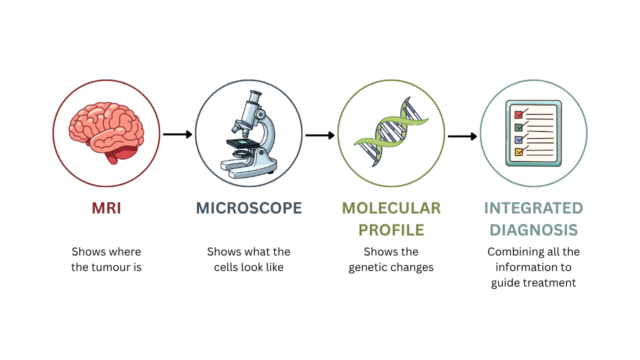
Markers such as IDH, 1p/19q, TERT, EGFR and CDKN2A/B can help determine what type of tumour a patient actually has.
MGMT is slightly different. It usually does not determine the name of the tumour, but it can help us understand how likely a glioblastoma is to benefit from temozolomide chemotherapy.
This is what makes modern brain tumour diagnosis so different from many other cancers
Clinical observation
Recently, I reviewed a patient who had been diagnosed with a diffuse glioma.
Interestingly, the MRI did not look like a typical aggressive brain cancer.
The tumour had poorly defined margins, which is common for gliomas because they grow by infiltrating normal brain tissue rather than forming a well-defined lump.
Even more surprising, there was no dramatic contrast enhancement, no obvious tumour necrosis and no imaging features that immediately suggested a highly aggressive cancer.
However, the molecular testing told a very different story.
The molecular results significantly changed how the tumour was classified and how we discussed prognosis and treatment.
What surprised me
One of the biggest surprises in modern neuro-oncology is this:
There are few cancers where molecular findings can become part of the diagnosis itself rather than simply helping us choose treatment.
In many cancers, such as breast, bowel or lung cancer, the pathologist first determines what the cancer is by examining the tissue.
Molecular testing may then help us select treatment.
Brain tumours can be different.
Under the current World Health Organization classification, many brain tumours now require doctors to combine the microscope findings with the tumour’s molecular results before giving the final diagnosis.
Sometimes a tumour may not look particularly aggressive on MRI or even under the microscope, but its molecular features tell us that biologically it belongs to a much more aggressive tumour group.
That is one of the most important changes in brain cancer diagnosis over the past decade.
Why brain cancer is different from most other cancers
Many cancers are described using stages:
- Stage I
- Stage II
- Stage III
- Stage IV
Gliomas generally are not classified that way.
Instead, doctors consider:
- what type of glioma it is;
- its CNS WHO grade; and
- its molecular profile.
For adult diffuse gliomas, doctors now recognise three main groups:
- astrocytoma, IDH-mutant;
- oligodendroglioma, IDH-mutant and 1p/19q-codeleted;
- glioblastoma, IDH-wildtype.
These names may sound complicated.
The important point for patients is that two tumours that look similar under the microscope may actually be different diseases once their DNA is tested.
Another important difference is that diffuse gliomas very rarely spread outside the central nervous system.
Unlike breast, bowel or lung cancer, the main problem is usually not spread to the liver, lungs or other organs.
Instead, the challenge is controlling tumour growth and infiltration within the brain itself.
Understanding glioma grades
Patients often see a WHO grade on their pathology report.
The grade is important, but it should no longer be interpreted on its own.
CNS WHO grade 1
Some grade 1 brain tumours, such as pilocytic astrocytoma, tend to grow slowly and are more circumscribed than adult diffuse gliomas.
When they can be completely and safely removed, surgery may sometimes be curative.
Grade 1 should not, however, be thought of as simply the first step in a pathway that inevitably progresses through grades 2, 3 and 4.
These can be biologically different tumour types.
CNS WHO grade 2
Grade 2 diffuse gliomas generally grow more slowly, but their cells infiltrate surrounding normal brain tissue.
Some may remain stable for many years.
Others can gradually become more aggressive over time.
CNS WHO grade 3
Grade 3 diffuse gliomas generally have more aggressive biological behaviour.
Treatment commonly involves surgery followed by additional treatment such as radiotherapy and chemotherapy, although the exact treatment depends heavily on the molecular diagnosis.
CNS WHO grade 4
Grade 4 represents the highest grade of adult diffuse glioma.
However, not every grade 4 glioma is called glioblastoma.
Today, the name glioblastoma is used for an IDH-wildtype tumour.
If the tumour has an IDH mutation, the diagnosis is instead:
Astrocytoma, IDH-mutant, CNS WHO grade 4.
In simple terms, two patients can both have a grade 4 glioma but still have biologically different tumours.
That difference can affect prognosis and treatment.
How can DNA change the diagnosis?
This is one of the most unusual parts of modern brain tumour diagnosis.
Sometimes a tumour does not look like a classic glioblastoma under the microscope.
However, if it is IDH-wildtype and has certain molecular changes, doctors may still classify it as glioblastoma.
These changes can include:
- a TERT promoter mutation;
- EGFR amplification; or
- a characteristic chromosome pattern called +7/−10.
You do not need to remember these names.
The important point is that the tumour’s DNA can sometimes reveal aggressive biology that is not obvious from the microscope alone.
There is a similar principle for IDH-mutant astrocytoma.
A change called CDKN2A/B homozygous deletion can make the tumour grade 4 even when some of the usual high-grade features are not seen.
This is why molecular profiling is now part of the diagnosis rather than simply an optional extra.
For many gliomas, it helps tell us what the tumour actually is.
What do the main molecular tests mean?
Modern glioma diagnosis may include several molecular markers. Some help define what type of tumour it is, while others help predict how the tumour may respond to treatment.
You do not need to remember every marker. The important point is that they answer different clinical questions.
| Molecular marker | What it helps tell us | Why it matters |
| IDH | Helps classify the type of adult diffuse glioma | Separates IDH-mutant astrocytoma from IDH-wildtype glioblastoma |
| 1p/19q codeletion | Helps identify oligodendroglioma | Oligodendroglioma requires both an IDH mutation and 1p/19q codeletion |
| TERT promoter mutation | Can support a glioblastoma diagnosis in the right IDH-wildtype tumour | May reveal aggressive tumour biology even when classic features are not obvious |
| EGFR amplification | Can support a glioblastoma diagnosis in the appropriate setting | Helps define certain IDH-wildtype diffuse astrocytic tumours as glioblastoma |
| CDKN2A/B deletion | Can affect the grade of an IDH-mutant astrocytoma | Homozygous deletion can establish CNS WHO grade 4 |
| MGMT promoter methylation | Helps estimate sensitivity to temozolomide | MGMT-methylated glioblastomas are generally more likely to benefit from temozolomide |
MGMT promoter methylation
What does MGMT tell us?
MGMT is different from the other markers.
It usually does not tell us what the tumour is.
Instead, it can help tell us how likely temozolomide chemotherapy is to work.
Why does MGMT matter?
MGMT is involved in repairing damaged DNA.
Temozolomide works partly by damaging DNA inside tumour cells.
When the MGMT promoter is methylated, the tumour is often less able to repair that damage, which means temozolomide may work better.
In the landmark study by Hegi and colleagues, about 45 in every 100 tumours tested were MGMT-methylated.
Among patients with MGMT-methylated tumours, median survival was approximately:
- 21.7 months with radiotherapy plus temozolomide
- compared with 15.3 months with radiotherapy alone.
These numbers describe groups of patients in a clinical trial. They should not be used to predict exactly how long one individual patient will live.
The important message is that MGMT helps us estimate how likely temozolomide is to help.
How is a glioma treated?
Treatment depends on:
- exactly what type of tumour it is;
- CNS WHO grade;
- molecular profile;
- the location of the tumour;
- neurological function;
- age;
- general health; and
- the individual patient’s goals and circumstances.
Whenever surgery can be performed safely, doctors usually aim for maximal safe resection.
This means:
remove as much tumour as possible, but not at the cost of causing serious permanent neurological damage.
The word safe matters.
Gliomas can infiltrate normal brain tissue, including areas responsible for speech, movement, memory or vision.
Removing every tumour cell is therefore often impossible.
The aim is to remove as much tumour as safely possible while avoiding permanent neurological harm. Current SNO/EANO guidance continues to emphasise maximal safe resection when feasible.
What happens after surgery for glioblastoma?
For many fit adults with newly diagnosed glioblastoma, a standard treatment approach is:
- radiotherapy;
- daily temozolomide during radiotherapy; and
- further cycles of temozolomide after radiotherapy.
This is commonly known as the Stupp regimen.
In the original Stupp trial, median survival improved from 12.1 months with radiotherapy alone to 14.6 months with radiotherapy plus temozolomide.
Another way to understand the benefit is to look at two-year survival:
- about 27 in every 100 patients receiving radiotherapy plus temozolomide were alive at two years;
- compared with about 10 in every 100 receiving radiotherapy alone.
These figures come from a study published in 2005 and should not be used to predict an individual patient’s outcome today.
They are extremely important historically, but they should not be used as an individual prognosis today because brain tumour classification, surgery, imaging and supportive care have evolved considerably since then.
For many fit adults with newly diagnosed glioblastoma, radiotherapy with concurrent and subsequent temozolomide nevertheless remains a standard first-line treatment approach.
What about older or frail patients?
Treatment does not have to be identical for everyone.
For older patients or those with significant medical problems or reduced functional status, shorter radiotherapy schedules may be appropriate.
Temozolomide may also be added depending on the patient’s condition and the molecular profile of the tumour.
In a trial of 562 patients aged 65 years or older, adding temozolomide to a shorter course of radiotherapy improved median survival from:
7.6 months to 9.3 months.
MGMT status can be particularly helpful when deciding how much benefit chemotherapy is likely to provide in this setting.
The important point is that age alone should not determine treatment.
We consider the patient’s overall health, neurological function, performance status, molecular findings and goals of care.
What does this mean for patients in Australia?
Modern glioma treatment usually requires several specialists working together.
Depending on the situation, this may include:
- neurosurgery;
- neuropathology;
- neuroradiology;
- radiation oncology;
- medical oncology;
- rehabilitation; and
- allied health support.
A multidisciplinary review is particularly important because the final diagnosis may depend on combining the MRI, surgery, pathology and molecular results.
In Australia, temozolomide is available through the Pharmaceutical Benefits Scheme as a Restricted Benefit, with multiple capsule strengths listed on the PBS.
Australian eviQ also provides endorsed protocols for temozolomide with radiotherapy followed by temozolomide in newly diagnosed glioblastoma, as well as shorter-course chemoradiotherapy approaches for selected older patients.
What would I usually consider?
When I review a patient with a newly diagnosed glioma, I try to answer a few important questions.
What does the MRI show?
Has the tumour been removed as safely and completely as possible?
What is the final diagnosis when we combine the microscope findings with the molecular results?
Is the tumour IDH-mutant or IDH-wildtype?
Is there a 1p/19q codeletion?
What does the MGMT result tell us about temozolomide?
How is the patient functioning neurologically?
Has the case been discussed by a specialist multidisciplinary team?
The important question is no longer simply:
“What does the tumour look like under the microscope?”
It is:
“What do all the pieces of information tell us when we put them together?”
The one message I want patients to remember
A brain tumour diagnosis today is much more than a name and a grade.
We may need to know whether the tumour is IDH-mutant or IDH-wildtype, whether it has a 1p/19q codeletion, and whether other molecular changes affect its diagnosis.
We may also use MGMT to help estimate how likely temozolomide is to work.
Patients do not need to memorise all of these terms.
The important point is that molecular testing can change the diagnosis, help us understand how the tumour may behave and guide treatment.
In modern neuro-oncology, the microscope tells us only part of the story. The molecular profile helps complete the picture.
Key take-home messages
- Brain tumours are classified differently from most other cancers.
- For many adult diffuse gliomas, molecular findings are now part of the diagnosis itself.
- Glioblastoma is now specifically an IDH-wildtype tumour; an IDH-mutant grade 4 tumour is classified as astrocytoma, IDH-mutant, CNS WHO grade 4.
- IDH and 1p/19q help define the tumour type, while MGMT is particularly useful for predicting likely benefit from temozolomide.
- Maximal safe surgery followed by appropriately selected radiotherapy and systemic treatment remains central to modern glioma care.
Key evidence
- Louis DN, Perry A, Wesseling P, et al. The 2021 WHO Classification of Tumors of the Central Nervous System: a summary. Neuro Oncol. 2021;23(8):1231-1251. doi:10.1093/neuonc/noab106
- Sahm F, Brandner S, Bertero L, et al. Molecular diagnostic tools for the World Health Organization (WHO) 2021 classification of gliomas, glioneuronal and neuronal tumors: an EANO guideline. Neuro Oncol. 2023;25(10):1731-1749. doi:10.1093/neuonc/noad100
- Wen PY, Weller M, Lee EQ, et al. Glioblastoma in adults: a Society for Neuro-Oncology (SNO) and European Society of Neuro-Oncology (EANO) consensus review on current management and future directions. Neuro Oncol. 2025;27(11):2751-2788. doi:10.1093/neuonc/noaf177
- Stupp R, Mason WP, van den Bent MJ, et al. Radiotherapy plus concomitant and adjuvant temozolomide for glioblastoma. N Engl J Med. 2005;352(10):987-996. doi:10.1056/NEJMoa043330
- Hegi ME, Diserens AC, Gorlia T, et al. MGMT gene silencing and benefit from temozolomide in glioblastoma. N Engl J Med. 2005;352(10):997-1003. doi:10.1056/NEJMoa043331
- Perry JR, Laperriere N, O’Callaghan CJ, et al. Short-course radiation plus temozolomide in elderly patients with glioblastoma. N Engl J Med. 2017;376(11):1027-1037. doi:10.1056/NEJMoa1611977
- Cancer Institute NSW. 3364-Glioblastoma temozolomide chemoradiation followed by temozolomide overview. eviQ. Accessed August 23, 2026. https://www.eviq.org.au/medical-oncology/neurological/glioma/3364-glioblastoma-temozolomide-chemoradiation-foll
Frequently asked questions
Does every brain tumour become glioblastoma?
No.
Many brain tumours never become glioblastoma.
Glioblastoma is now a specific molecularly defined adult diffuse glioma that is IDH-wildtype.
Is every grade 4 brain tumour a glioblastoma?
No.
This is an important change in modern terminology.
Glioblastoma is CNS WHO grade 4 and IDH-wildtype.
An IDH-mutant grade 4 diffuse astrocytic tumour is called astrocytoma, IDH-mutant, CNS WHO grade 4 rather than glioblastoma.
What is an IDH mutation?
IDH is one of the most important molecular markers used to classify adult diffuse gliomas.
It helps separate biologically different tumour types and provides important prognostic information.
What does 1p/19q codeletion mean?
A tumour must be both IDH-mutant and 1p/19q-codeleted to be classified as an oligodendroglioma under the current WHO system.
Can molecular testing really change my diagnosis?
Yes.
For many gliomas, molecular findings are now essential diagnostic criteria.
In some situations, they can change both the final tumour name and its grade.
What does MGMT methylation mean?
MGMT promoter methylation is associated with a greater likelihood of benefit from temozolomide in glioblastoma.
It does not usually define what type of glioma you have.
Why don’t gliomas usually spread to the liver or lungs?
Diffuse gliomas very rarely spread outside the central nervous system.
Their main challenge is usually growth and infiltration within the brain itself.
Is surgery always possible?
No.
Some tumours involve areas responsible for functions such as speech, movement or vision.
The neurosurgical team balances removing as much tumour as possible against the risk of causing permanent neurological injury.
Can surgery cure glioblastoma?
Surgery is extremely important, but glioblastoma infiltrates surrounding brain tissue.
For this reason, surgery alone is generally not curative and is usually followed by additional treatment.
What is the Stupp regimen?
The Stupp regimen combines radiotherapy with daily temozolomide followed by further cycles of temozolomide.
For many fit adults with newly diagnosed glioblastoma, it remains a standard first-line treatment approach.
About Dr Dilanka De Silva
Dr Dilanka De Silva is a Melbourne Medical Oncologist and Cancer Genetics Physician with an interest in precision oncology and complex cancer second opinions.
He completed advanced fellowship training at Memorial Sloan Kettering Cancer Center in New York.
Dr Dilanka provides consultations and second opinions for patients with brain tumours and gliomas in Melbourne and also reviews international patients from Sri Lanka, the Maldives, Indonesia and the Philippines.
Where appropriate, treatment planning can be coordinated with the patient’s neurosurgical, radiation oncology and local oncology teams.
Dr Dilanka De Silva
MBBS, MRCP(UK), MRCP(UK SCE Oncology), FRACP, PhD
Medical Oncologist & Cancer Genetics Physician
Memorial Sloan Kettering Cancer Center Fellow (New York, USA)